There are many reasons to have an eye exam. Vision correction, of course, but also detection of eye disorders like glaucoma and macular degeneration – many of which can be slowed or even stopped if diagnosed early.1 But did you know that an eye exam is often the first to diagnose serious health conditions like diabetes, high cholesterol and high blood pressure?2
Now that you’re convinced to get an eye exam, what should you expect during your visit? The first thing to know is that a vision exam is more involved than a simple vision screening. Below, we walk you through some of the standard steps of a vision care exam and explain why each is important.
Preparing for the exam
If you wear glasses or contacts, be sure to bring them to the exam. Your eye doctor will want to examine your vision with and without your corrective lenses. Also, you’ll want a pair of sunglasses to help you avoid bright lights in case your eyes have been dilated during the exam. Dilation can last several hours and may cause light sensitivity and blurry vision, so you may want to consider having someone drive you home.
During the exam
A technician may perform some of the initial tests and provide the results to the eye doctor for review.
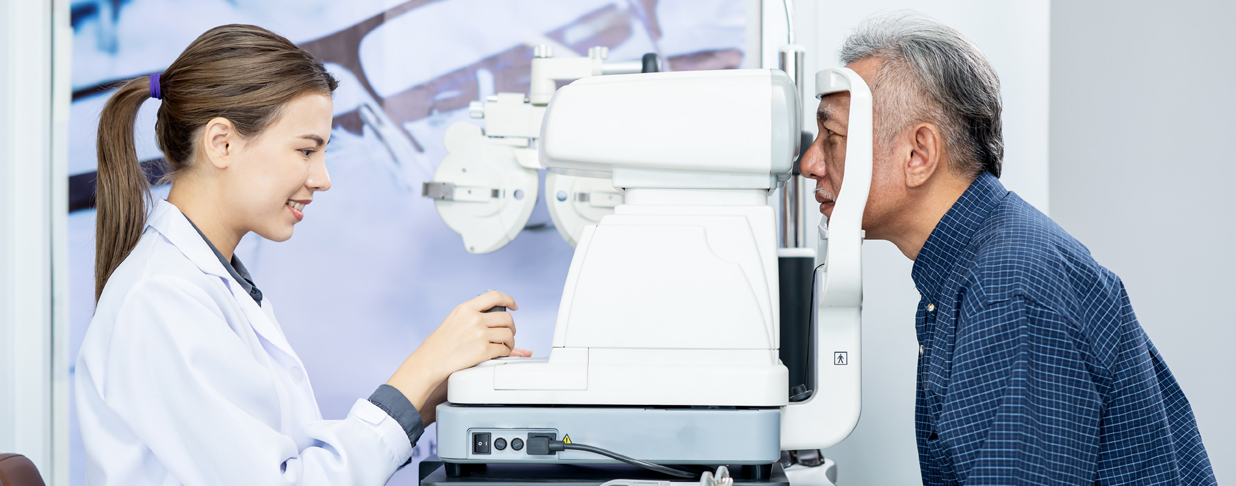
Glaucoma test
The most common version of the glaucoma test is also known as the “puff-of-air” test, though it is formally called non-contact tonometry. After putting your chin on the instrument’s chin rest, you’ll look at a light while a small burst of air is directed at your eye. It’s common to flinch, but know that this process is painless and the tonometer never touches your eye.
Your eyes’ resistance to the puff of air indicates the pressure inside your eye. If the pressure is high, you may be at risk for glaucoma and require further testing.3
There are also other instruments to measure the pressure that may be used which painlessly tap the surface of the eye for a very brief instant.
Visual field test
The visual field test determines the fullness of your field of vision and identifies blind spots in your peripheral vision. Knowing if there are areas with limited vision can help your eye doctor diagnose certain eye conditions.3
The doctor will perform the test either manually by asking you when you see their hand move into your peripheral vision, or by using an automated perimetry instrument. With the automated test you will be directed to sit and look into a bowl-shaped device. While you stare at the center of the bowl, lights flash randomly in various locations. You press a button every time you see a light flash.
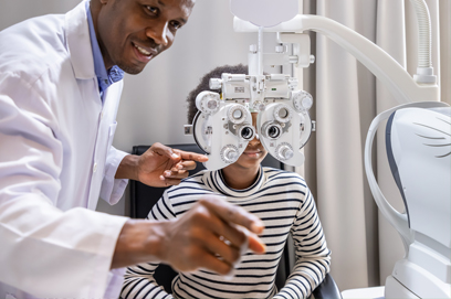
Refraction
This test allows the doctor to maximize your visual acuity by putting a mask-like instrument called a phoropter in front of your eyes.
You will see an eye chart through a series of corrective lenses and the doctor will ask you which lens options look clearer. You may be familiar with the standard “which looks better, 1 or 2” question.
Your answers will help the eye doctor fine tune the lens power until reaching a final prescription that works best for your vision.
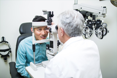
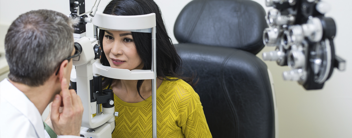
Slit lamp test
A slit lamp is essentially a microscope for your eyes. Using this device, the eye doctor can examine your eyelids, tears, cornea, iris and lenses. A dye (fluorescein) may be applied to your eyes to help reveal damaged cells. Tears wash the dye away quickly, so it won’t be noticeable by the time you leave the exam.
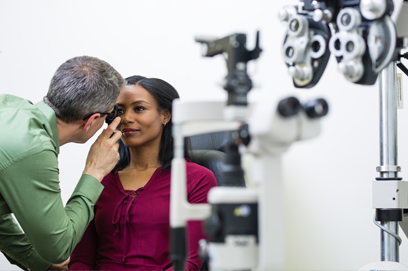
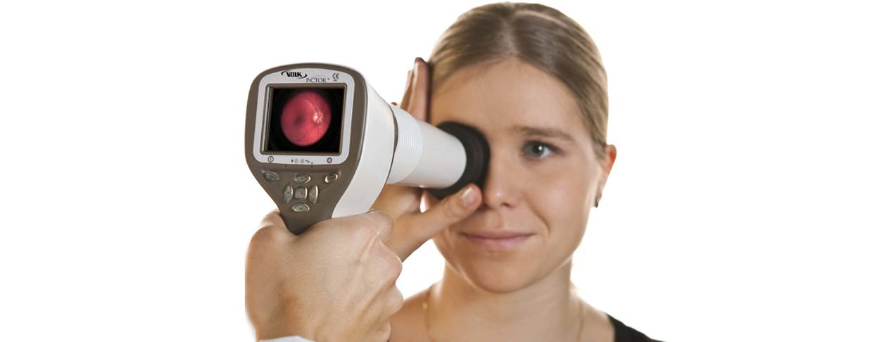
Retinal viewing test
Also called ophthalmoscopy, this retinal viewing test provides the eye doctor an in-depth view of the back of your eye, the optic disk and the retinal blood vessels.
Dilation drops are often used to obtain a better view. Dilation requires 20 minutes to take effect, so your eye doctor may apply the drops early in your vision exam. After your pupils are dilated you will experience light sensitivity and difficulty focusing on close objects. These effects may last for several hours, so plan accordingly.
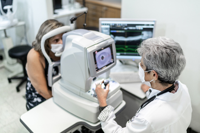
Once your eyes are fully dilated, the doctor will shine a beam of light through your pupil and exam the retina and the back of your eye. This unobstructed view of your small blood vessels is also what allows the doctor to identify early stages of diabetes, high cholesterol and high blood pressure.2 If any of these conditions are observed, your eye doctor will record the findings and refer you to your primary care provider or other specialist for further testing.
An alternative to pupil dilation includes retinal imaging devices that provide high-resolution photos of your retina. The doctor will save these images and refer to them for subtle retinal changes over time. When scheduling your appointment, ask if this technology is available or if you’ll need pupil dilation.
Exam results
A vision care exam will determine if you have vision correction needs and your doctor will provide lens prescriptions for either glasses or contacts. Your eye doctor may also discuss whether you’re a candidate for vision correction surgery.
The exam will also reveal any eye health concerns such as cataracts, glaucoma or retinal disorders, such as macular degeneration or diabetic retinopathy. Early detection of these disorders are key to stopping their progression and preserving vision.1
To learn more about the importance of an annual eye exam and how EyeMed removes the barriers to getting one, contact your EyeMed representative or visit eyemed.com.
••••
1 - “Avoid Vision Loss By Catching Conditions Early,” yoursightmatters.com, accessed April 2020.
2 - MedCity News - “Comprehensive managed vision care is more than mere “medical management,” Klunk, E., Sept 20, 2019,
3 - “Eye Exam," mayoclinic.org, accessed October 2021.