In our latest interview with EyeMed’s medical team, Dr. Lahr and Dr. Neighbors, we discuss retinal imaging and its benefits. Here’s what we learned.
"We get lots of questions about retinal imaging. What is it? Why is it so important? And why isn’t it covered as part of an eye exam?”
Your eye is only about 1 inch across. Yet it’s one of the most complex organs in your body. To help providers see your inner eye more clearly, a retinal image can be critical.
A retinal image sees up to 5 times more than a traditional undilated eye exam. Plus, a retinal image is an important historical baseline that allows providers to compare images year-over-year so that conditions can be managed and treated.
What exactly is a retinal image?
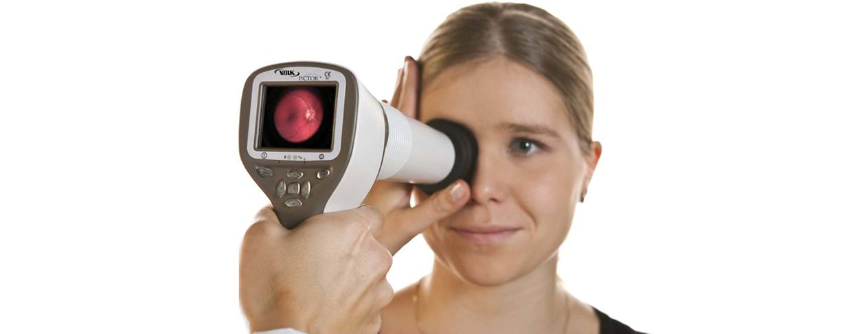
Retinal imaging is a high-resolution image of the inside of your eye. Included in the image is a detailed view of the retina, optic nerve and retinal blood vessels—all of which can show signs of underlying eye and general health issues.
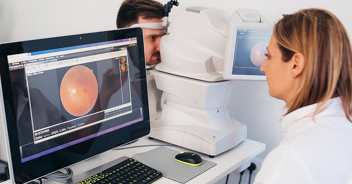
How is a retinal image captured?
A retinal image is quick and painless. The patient sits comfortably in a chair and places their forehead and chin on the camera device. Nothing touches the eye during retinal imaging. A quick flash of light, and the image is captured in real-time.
What can be diagnosed through a retinal image?
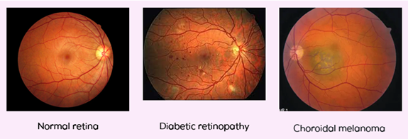
Retinal imaging is a non-intrusive way to see the early signs of vision and health conditions:
- Cataracts
- Glaucoma
- Diabetic retinopathy
- Macular degeneration
- Some forms of eye cancer
- Diabetes
- Hypertension
- High cholesterol
Do you need pupil dilation with retinal imaging?
Pupil dilation allows observation of the peripheral retina. However, dilation is not always convenient for patients due to the side effects— blurry vision and light sensitivity—for several hours after the exam. Retinal imaging allows the same view of the peripheral retina without pupil dilation and the undesired side effects.
So why can’t retinal imaging be added to the components of a comprehensive eye exam and avoid the added out-of-pocket cost?
What would seem simple is actually very difficult and challenging. Eye exam service code definitions, which include the test components, are controlled through copyright by the American Medical Association. If you think new codes are easy to create, think again.
EyeMed introduced Retinal Imaging 7 years ago and the popularity of this screening test has continued to grow. Currently, the median acceptance rate for Retinal Imaging is 65%.
Meet the experts: Medical Directors, Drs. Lahr and Neighbors

Dr. John Lahr, O.D., FAAO Dr. Andrew Neighbors, O.D.
For more information, reach out to your EyeMed representative or visit eyemed.com.